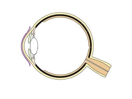
43 diagram of the human eye without labels
Eye Quizlet Labeling Search: Eye Labeling Quizlet. Zygomatic Bone 13 The 6 to 7 million cones provide the eye's color sensitivity and they are much more concentrated in the central yellow spot known as the macula Select different colors for the bone regions listed at the coding circles below (Label your answer with the proper units Ear part identification and labeling for labratory test learn with flashcards games ... The Body Parts in Spanish - From Head to Toe - Mondly Blog Here are all the body parts you can find in this region: (the) brain — (el) cerebro (the) face — (la) cara (the) hair — (el pelo) (the) forehead — ( la) frente (the) cheek — ( la) mejilla (the) ear — (la) oreja (the inside ear is ' el oído') (the) eye — (el) ojo (the) eyelid — (el) párpado (the) eyelashes — ( las) pestañas
Anatomical Position and Directional Terms - EZmed We will first review the anatomical position, its definition, and look at example labeled diagrams. We will then walk through the different anatomical directional terms used to describe location and movement. We will provide you with labeled diagrams, example body parts, and tricks to learn the directional terms listed below! Medial vs Lateral

Diagram of the human eye without labels
What are the 12 cranial nerves? Functions and diagram The cranial nerves are a set of twelve nerves that originate in the brain. Each has a different function responsible for sense or movement. The functions of the cranial nerves are sensory, motor ... Revealing the human mucinome | Nature Communications F Venn diagram comparing mucinomics results from five cell lines. Each sample is shown as a different color (red, orange, purple, green, and blue); 26 mucin proteins were enriched in all five samples. anatomydiagram123.z21.web.core.windows.netdiagram of human eye Human Sense Organs - The Five Senses . eye sense organs human sight structure organ senses vision five eyes ojo brain anatomy. Bionic Eye 5 - Innovate Long Island . eye bionic eyes intelligence future blind artificial chip implant tell pink vision cyborg fix reflections ecosystems woman sight october ...
Diagram of the human eye without labels. Anatomical Position: Body Planes and Sections - EZmed The transverse plane is the only horizontal plane, and it divides the body into top (superior) and bottom (inferior) sections. An easy trick to remember the transverse plane is to again use the name. The "Transverse" plane will give you a "Top View" of the body as it divides the body into upper and lower portions. Corneal Reflex - StatPearls - NCBI Bookshelf The cornea is a smooth, clear structure at the front of the eye. It functions to (1) shield the eye from foreign substances and (2) help control visual focus.[1][2] To focus light, the cornea must be clear; therefore, it has no blood vessels to impede light refraction. Tears and the aqueous humor of the eye nourish it (fluid in the anterior part of the eye between the cornea and the pupil and ... The 14 Facial Bones: Anatomy & Functions - Study.com Here is a labeled diagram of the fourteen facial bones: Facial Bone Names Let's begin with the nasal bones. In this case, we're actually looking at two small bones, which are located just above the... Ear Diagram Quiz - ProProfs Ear Diagram Quiz 14 Questions | By Bellamiller123 | Last updated: Mar 22, 2022 | Total Attempts: 6037 Questions All questions 5 questions 6 questions 7 questions 8 questions 9 questions 10 questions 11 questions 12 questions 13 questions 14 questions
Quiz: Label The Parts Of The Eye - ProProfs How much did you get to understand about the human eye? Take up this quiz and find out! Questions and Answers. 1. A is pointing to what part of the eye? A. Cornea. B. Optic Nerve. WHMIS 2015 - Pictograms : OSH Answers With a quick glance, you can see, for example, that the product is flammable, or if it might be a health hazard. Most pictograms have a distinctive red "square set on one of its points" border. Inside this border is a symbol that represents the potential hazard (e.g., fire, health hazard, corrosive, etc.). › en › libraryAnatomy of the eye: Quizzes and diagrams - Kenhub Oct 28, 2021 · Take a look at the diagram of the eyeball above. Here you can see all of the main structures in this area. Spend some time reviewing the name and location of each one, then try to label the eye yourself - without peeking! - using the eye diagram (blank) below. Unlabeled diagram of the eye. Click below to download our free unlabeled diagram of ... Anatomical Planes of Body - The Human Memory The X-axis is going from left to. right, Z-axis from front to back, and Y-axis from up to down. In anatomical. terminology, three references plane are considered standard planes; these. planes differentiate the body anterior and posterior, ventral and dorsal, dexter, and sinister portions. Let me tell you about these standard planes in detail.
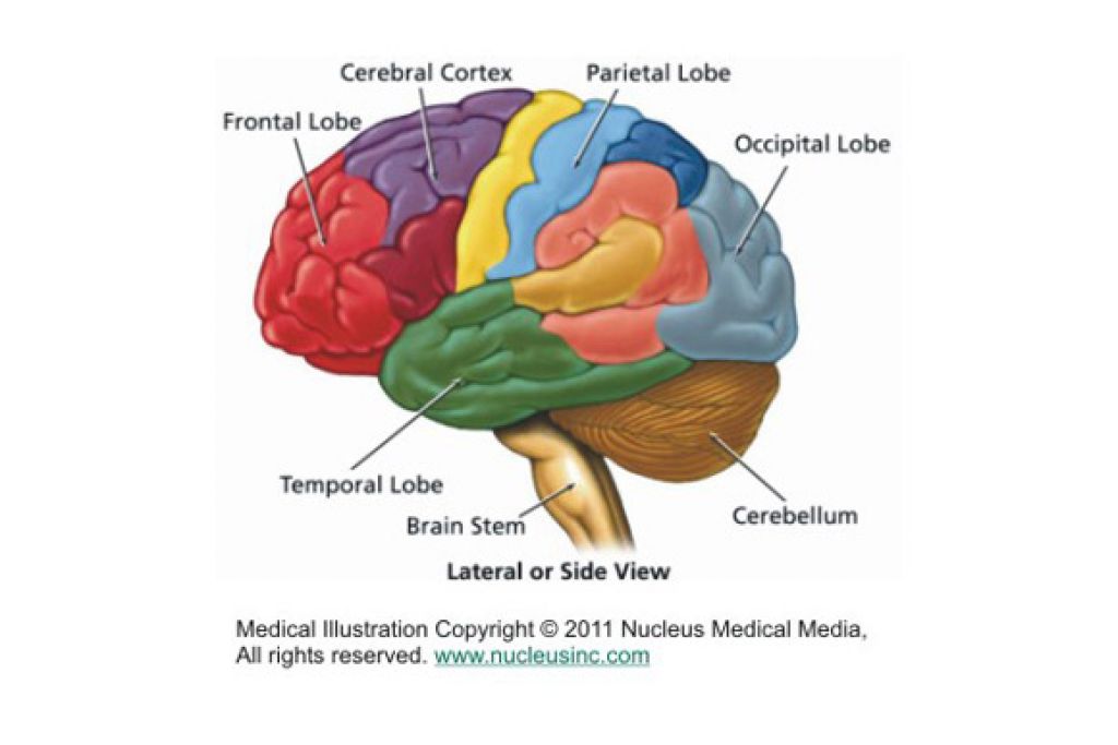
Blank ear diagrams and quizzes: The fastest way to learn - Kenhub It helps you to memorize the names and their locations, which in turn will aid you to remember their functions. Below, you can download both the blank ear diagram to make some notes, and then try labeling the ear using the unlabeled ear diagram. Good luck! DOWNLOAD PDF WORKSHEET (BLANK) DOWNLOAD PDF WORKSHEET (LABELED) CBSE Class 10 Science Important Biology Diagrams For Last Minute ... CBSE Class 10 Chemistry Important Reactions. 2. Brain. A human brain is composed of three main parts- the forebrain, the midbrain and the hindbrain. These three parts have specific functions ... Free Nervous System Worksheets and Printables The human nervous system is the part of human anatomy that sends signals from sensory receptors to the spinal cord and brain and then transmits the impulses back to the other parts of the body. ... Label the Brain Anatomy Diagram - This brain anatomy labeling worksheets is a great addition to the study of human anatomy. Conjunctiva: Anatomy, Function, and Treatment - Verywell Health Function. The primary function of the conjunctiva is to keep the front surface of the eye moist and lubricated. It also keeps the inner surface of the eyelids moist and lubricated, making them able to open and close easily without causing eye irritation. Another job of the conjunctiva is to protect the eye from dust, debris, and microorganisms ...

Human Body Anatomy Basics No Lines Clip Art at Clker.com - vector clip art online, royalty free ...
commons.wikimedia.org › wiki › File:SchematicFile:Schematic diagram of the human eye no.svg - Wikimedia Mar 28, 2022 · Original upload log []. This image is a derivative work of the following images: File:Schematic diagram of the human eye en.svg licensed with PD-self 2008-02-02T01:33:45Z Jakov 508x516 (54267 Bytes) suspensory ligament, arrow was wrong

Human Skeleton Blank Clip Art at Clker.com - vector clip art online, royalty free & public domain
Simple Microscope - Parts, Functions, Diagram and Labelling Parts of the optical parts are as follows: Mirror - A simple microscope has a plano-convex mirror and its primary function is to focus the surrounding light on the object being examined. Lens - The biconvex lens is placed above the stage and its function is to magnify the size of the object being examined.
picture front of the eye without labels clipart - Clipground
Macular hole morphology and measurement using an automated three ... A. Schematic diagram of the macula hole 3D model; B. OCT of a macula hole with 2D labels; C. Representative example of a segmented 3D macular hole in 3 different orientations - base area marked by *, top area (i.e. at the ILM side) marked by #. ... the preoperative visions were checked without a protocol refraction and exact relationships are ...
Free Brain Diagram, Download Free Brain Diagram png images, Free ClipArts on Clipart Library
Anatomy of the Eye | BrightFocus Foundation Optic nerve: The bundle of nerve fibers at the back of the eye that carry visual messages from the retina to the brain. Photoreceptors: The light sensing nerve cells (rods and cones) located in the retina. Pupil: The adjustable opening at the center of the iris through which light enters the eye. Retina: The light sensitive layer of tissue that ...

Eye - Labelled Diagram Of Human Eye | Transparent PNG Download #836865 - Vippng
Anatomical diagrams of the brain - e-Anatomy - IMAIOS Atlas of the human brain based on colored anatomical drawings and diagrams. ... The basic structure of a neuron and an overall diagram of the human nervous system. Meninges : Coronal section . ... The user can select to display multiple categories of labels on the illustrations: Cerebral lobes / regions; Cerebrum, divided into: frontal lobe ...

picture front of the eye without labels clipart 20 free Cliparts | Download images on Clipground ...
Brigitte Zimmer The human heart is one of the most essential parts of the human body, and getting to know it is imp… Read more How To Draw Human Heart And Label Outline Human Heart Diagram Class 10
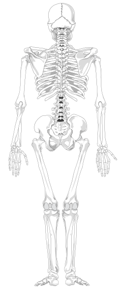
Human Skeleton Back No Text No Color Clip Art at Clker.com - vector clip art online, royalty ...
› GAM2012_Eye_Diagram_HandoutParts of the Eye - National Institutes of Health Eye Diagram Handout Author: National Eye Health Education Program of the National Eye Institute, National Institutes of Health Subject: Handout illustrating parts of the eye Keywords: parts of the eye, eye diagram, vitreous gel, iris, cornea, pupil, lens, optic nerve, macula, retina Created Date: 12/16/2011 12:39:09 PM

#89 Structure and function of the eye, rods and cones | Biology Notes for IGCSE 2014
Parts of the Microscope with Labeling (also Free Printouts) 5. Knobs (fine and coarse) By adjusting the knob, you can adjust the focus of the microscope. The majority of the microscope models today have the knobs mounted on the same part of the device. Image 5: The circled parts of the microscope are the fine and coarse adjustment knobs. Picture Source: bp.blogspot.com.
Post a Comment for "43 diagram of the human eye without labels"